Greek Origins of Animal Classification
The Classification of Urochordate
The animals which have a multicellular are usually divided to two groups vertebrates and invertebrates and this segmentation back to 500 years BC (Satoh et al,2014). In the ancient Greek civilization Aristotle 300 years BC recognised the animal based on blood to (Enaima with blood) and (Anaima without blood) (Satoh et al,2014). According to Satoh et al, (2014) when Darwin established the evaluation of animals the scientist investigates in more than one century (Satoh et al,2014). This classification still continued until Linnaeus find the new taxonomy system and Carolus Linnaeus was a botanist who is divided the animal and plant in different group (Satoh et al,2014). The phylum of chordate divided into three different groups: urochordate, cephalochordate and vertebrates (Satoh et al,2014) (Cole,2012). The amphioxus is the animal of the cephalochordate group and this name is a result to the notochord is cover the all anterior tip (Nishino and Satoh,2011). Therefore, another subphylum belongs to the chordate phylum is called Urochordata (tunicate) (Nishino and Satoh,2011). This subphylum has five groups appendicularia (Larvacea), ascidiacea, pyrosomida, doliolida and salpida (Nishino and Satoh,2011). Ascidians are the biggest group in the subphylum urochordate (Hirose,2009). Ali & Tamilselvi (2016) suggest that the classification of tunicate based on the morphological structure. This essay will indicate the subphylum urochordate based on morphology, taxonomy, classification and lifestyle of urochordate specially ascidians.

Class ascidiacea:
Ascidians are the biggest class in the subphylum Urochordata because has more than 2300 species which are discovered (Hirose,2009). Ali and Tamilselvi (2016) suggest that this class including more than 3000 species around the world. According to McHenry and Patek (2004), ascidians are the huge number of the marine invertebrate with more than 3000 species. This class follows the phylum chordate because the larvae stage has a notochord (McHenry and Patek,2004). This class has a massive number of families under two orders: Enterogona and Pleurogona (Ali and Tamilselvi,2016). Ascidiacea class has lifestyle such as free swimming in larva stage and also has two stages in the circle life sometimes lives as an individual and another times lives as a colony (Ali and Tamilselvi,2016) (Cole,2012). The organisms follow this class which life as a colony reach to two meters long, however, the length of others life as an individually from one millimetre to 20 centimetres (Britannica.com,2016). Additionally, class ascidiacea has two siphons are pointed upward in the direction (Ali and Tamilselvi,2016).
Order: Enterogona
The organisms which follow this order have simple body structure and lives in as a colony (Ali and Tamilselvi,2016). The body style of this order divided to two parts thorax and abdomen also, this order has a simple tentacle, beside the reproductive system opens in the anus and follow the rectum (Ali and Tamilselvi,2016). Actually, in this order found the gland of the neural system on the ventral side (Ali and Tamilselvi,2016). This order contains two suborders Aplousobranchiata lahille, phlebobranchiata lahille (Ali and Tamilselvi,2016). The first suborder is Aplousobranchiata lahille and this suborder founded as colony ascidians. Zooids extend to all body and divided to thorax and abdomen (Ali and Tamilselvi,2016). Moreover, the post abdomen could be found in this suborder also, the branchial sac has a long inner vessel, simple, transverse (Ali and Tamilselvi,2016). The gut canal is founded in abdomen and gonads in the abdomen side (Ali and Tamilselvi,2016). This suborder has three families: Clavelinidae Forbes, Polyclinidea Milne and Didemnide (Ali and Tamilselvi,2016). This first family is Clavelinidae Forbes, this family including the colony and the ascidia in this family lives as a communal (Ali and Tamilselvi,2016). The body is divided to thorax and abdomen furthermore, the gill in this family more than three slits (Ali and Tamilselvi,2016). The gonads in this family inside the intestine and “atrial siphon opens to extensive directly or into a common cloace” (Ali and Tamilselvi,2016). “True post abdomen is absent” (Ali and Tamilselvi,2016). This family has three subfamilies Polycitorinae, Clavelininae and Holozoinae (Ali and Tamilselvi,2016).
The first subfamily Polycitorinae and the characteristic of this subfamily the atrial and branchial siphon have six lobed (Ali and Tamilselvi,2016). Moreover, the reproductive system in a sexual way by budding from the abdomen side (Ali and Tamilselvi,2016). This subfamily including several genera such as Polycitor, Eudistoma, Archidistoma, Tetrazoma, Cystodytes and Sigillina (Ali and Tamilselvi,2016). Clavelininae is the second subfamily and they live as a colony and individual forms, the siphons are open to the external way, on the other hand, the branchial vessels are absent (Ali and Tamilselvi,2016). Clavelininae has five genera Clavelina, Podoclavelina, Pycnoclavelina, Archiascidia, Oxycorynia (Ali and Tamilselvi,2016). The third subfamily under this family is Holozoniae, this subfamily has six lobed in the branchial siphon “whereas atrial siphons are the modified atrial languets. Brood pouch with embryos present at thoracic level. Buds are produced at end of the abdomen” (Ali and Tamilselvi,2016). This subfamily has Distaplia, Sycozoa, Corella and Holozoa as genera (Ali and Tamilselvi,2016). Ali and Tamilselvi (2016) indicates that the second family in this suborder is Polyclinidae Milne. This family live as a colonial ascidian also, this family often are a huge and gelatinous (Ali and Tamilselvi,2016). This family has seven or more than seven rows of stigmata in the huge zooids, “Branchial aperture is lobed, whereas atrial aperture is not lobed. Atrial siphon opens into common cloaca. Body of the zooid is divided into thorax, abdomen, posterior abdomen. Gonads and heart are present in post abdomen. Budding is by post abdominal constrictions” (Ali and Tamilselvi,2016). The family Polyclinidae contains two subfamilies Polyclininae and Euherdmaniae (Ali and Tamilselvi,2016. The first subfamily is Polyclininae has “Polyclinids with atrial apertures forming common cloaca” (Ali and Tamilselvi,2016). in addition, this subfamily has between six and eight lobed in the branchial siphon this caused to divided this subfamily to four parts, the first part which has six lobed in the branchial siphon and the wall of stomach is smooth and this include the Polyclinum, Aplidiopsis, Sydneioides genera (Ali and Tamilselvi,2016). On the other hand, the part which has six lobed in the branchial siphon and the stomach with ridges has three genera like Aplidium, Amaroeucium, Synoicum (Ali and Tamilselvi,2016). The genera Sydnyum and Morchellium have eight lobed in the branchial siphon and also the stomach with ridges The last part is called aberrant has Pharyngodictyon (Ali and Tamilselvi,2016). In contrast, the subfamily Euherdmaniae has a free atrial siphon and has Euherdmaniae (Ali and Tamilselvi,2016). The third family in this suborder is Didemnidae, the scientist found this family in a colonial form (Ali and Tamilselvi,2016). Colonies have covered by a hard surface layer and the zooids are divided between thorax and abdomen also the zooids are very short (Ali and Tamilselvi,2016). Atrial siphons have changed the form to atrial lip, moreover, in this family is clear to see the calcareous spicules also, the gonads in the dorsal posterior, “budding is complex involving epicardia” (Ali and Tamilselvi,2016). This family covered the several genera such as Didemnum, Trididemnum, Diplosoma, Leptoclinides, Lissoclinum, Echinoclinum, Leptoclinum, Coelocarmus . (Ali and Tamilselvi,2016).
The second suborder in this order is Phlebobranchiata Lahille (Ali and Tamilselvi,2016). This suborder lives in both lifestyle (colonies or individually) in the family called Diazonidae Garstang the body does not divide to thorax and abdomen, “Branchial sac is with internal longitudinal vessels or in the form of bifurcating branchial papillae, but never with folded. Gonads are present in gut loop or at the side of branchial sac”. Phlebobranchiata contains seven families (Ali and Tamilselvi,2016). The first family is Cionidae, this family lives as an individual style (Ali and Tamilselvi,2016). In the first time the digestive tube is clear in this family and in the horizontal feature and in posterior to the thorax (Ali and Tamilselvi,2016). Branchial sac has several rows of stigmata and this branchial is huge also the heart in V-shaped and is located between stomach and endostyle (Ali and Tamilselvi,2016). “inner longitudinal vessels with secondary papillae” (Ali and Tamilselvi,2016). Gonads found in the gut loop and both of them (oviduct and spermduct) open near to the anus, this family includes genus Ciona (Ali and Tamilselvi,2016). The second family is Diazonudae , this family has both situations to lives, sometimes lives as colonial and another time lives as an individual form (Ali and Tamilselvi,2016). Also, all zooid has a separate branchial siphon and atrial, moreover, the body is divided to thorax and abdomen (Ali and Tamilselvi,2016). Gut loop and gonads are together and open near the anus, the branchial sac in rows of stigmata with numerous also, inner longitudinal vessels are founded with primary papillae, however, secondary papillae are disappeared (Ali and Tamilselvi,2016). The genera including this family are Diazona, Tylobranchion, Rhopalea (Ali and Tamilselvi,2016). The next family under this order is Perophoridae, the branchial sac in the gut loop side the gut loop (Ali and Tamilselvi,2016). “gut loop is lower” (Ali and Tamilselvi,2016). “The branchial sac with longitudinal vessel is in the form of bifurcating primary papillae. Secondary papillae are absent. The heart is long up to the base of endostyle along the dorsal side. Epicardia are absent” (Ali and Tamilselvi,2016). Gonads located in the gut loop also, sperm duct opens in the anus, however, the oviduct is short and opens into matrix cavity (Ali and Tamilselvi,2016). The most genera under this family Perophora, Ecteinascidia (Ali and Tamilselvi,2016). The fourth family including this order is Corellidae Lahille this family lives as an individual solitary gut loop is near to the end of the body and located on the right side regards branchial sac (Ali and Tamilselvi,2016). Gonads near to the anus and located in the gut loop and branchial sac dose not have secondary papillae, however, has inner longitudional vessels (Ali and Tamilselvi,2016). This family has several genera such as Corella, Rhodosoma, however, some genus is imperfectly known (Ali and Tamilselvi,2016). The fifth family under this suborder is Ascidiidae this family has lifestyle as individual forms, the only point different with the previous family is the gut loop, in this family, is on the left side on branchial sac (Ali and Tamilselvi,2016). On the other hand, the similar point with Corellidae Lahille family gonads are located near to the anus and in the gut loop (Ali and Tamilselvi,2016). “the branchial sac has inner longitudinal vessels, with or without secondary papillae also, the epicardia are represented by mumerous renal vesicles” (Ali and Tamilselvi,2016). This family has three genera, Ascidia, Ascidiella and phallusia (Ali and Tamilselvi,2016). The last two families in this suborder are Hypobythiidae and Agnesiidae, the genera in these family are live in highly deep water respectively, therefore, all organisms in these family are related to Ascidiidae (Ali and Tamilselvi,2016).
Order: Pleurogona
In this order found the both forms colony and simple also, the body is one part without divided to thorax and abdomen (Ali and Tamilselvi,2016). Gonads are located on the lateral side on the wall and both sides of the body (Ali and Tamilselvi,2016). In the dorsal side, this order usually has neural gland and sometimes lateral to ganglion (Ali and Tamilselvi,2016). Moreover, the tentacles could be compound or simple and buds are created from the mental wall (Ali and Tamilselvi,2016). This order includes one suborder Stolidobranchiata this suborder lives as a colony or single animal, branchial sac with inner longitudinal vessels or bars (Ali and Tamilselvi,2016). This suborder has three families. This first family is Styelinae. This family stays in as single animal or as a colony style like ascidians (Ali and Tamilselvi,2016). This family has two siphons and both of them are smooth edged also each siphon has four-lobed (Ali and Tamilselvi,2016). Branchial sac contains four folds in the both sides (Ali and Tamilselvi,2016). “dorsal lamina has smooth-edged” (Ali and Tamilselvi,2016). The structure of tentacles is very simple and the “stomach is wide ridges and pyloric caecum” (Ali and Tamilselvi,2016). This family has two subfamilies. The first one is Styelinae. This subfamily found as a single animal and has two siphons both of them has four-lobed (Ali and Tamilselvi,2016). Branchial sac normally has four folds on both sides (Ali and Tamilselvi,2016). This subfamily has two genera based on the location of gonads if the gonads are in one side or both sides that means include Pelonia, Styela, Katatropa, Polycarpa and Cnemidocarpa (Ali and Tamilselvi,2016). In contrast, gonads in this genera Dendrodoa, Podostyela are just in the one side (Ali and Tamilselvi,2016). Second subfamily which is lives as a colony is Botryllinae and these colonies either join with zooids or not (Ali and Tamilselvi,2016). The mantle wall generated the buds (Ali and Tamilselvi,2016). Oviduct and spermduct are changeable, the larvae of this subfamily have a special sensor called “photolith” (Ali and Tamilselvi,2016). This subfamily has two genera based on branchial sac is with folds or without folds, the first genera include Polyandrocarpa, Eusynstyela, Gynandrocarpa, Stolonica and Distomus have “branchial sac with folds and numerous inner longitudinal vessels or bars” (Ali and Tamilselvi,2016). The second genera in this subfamily have branchial sac without folds but with the part which is residual from longitudinal vessels or bars (Ali and Tamilselvi,2016). The second family which has two siphons and each of them has four-lobed also tentacles are branched (Ali and Tamilselvi,2016). Branchial sac located in both sides and actually has four folds in each side (Ali and Tamilselvi,2016). A great number of internal longitudinal vessels found in this family and stigmata are straight (Ali and Tamilselvi,2016). The stomach in this family is tight and smooth (Ali and Tamilselvi,2016). This family called Pyuridae and has a several genera such as Pyura, Boltenia, Microcosmus, Tethyum and Culeolus (Ali and Tamilselvi,2016). The last family in this suborder is Molgulidae. According to Ali and Tamilselvi (2016), this family has two siphons as well but the different with the previous family is the number of lobed because this family has six lobed. As well as tentacles are branched (Ali and Tamilselvi,2016). In this family founded six or seven branchial folds with numerous longitudinal vessels in the branchial sac (Ali and Tamilselvi,2016). The next characteristic of this family is the cheeped for stigmata is turned and arranged around spirals (Ali and Tamilselvi,2016). In the left side founded the kidney near to the sac and is a large organ (Ali and Tamilselvi,2016). The genera follow this family are Molgula and Eugyra (Ali and Tamilselvi,2016).
Morphology of Ascidians:
The epidermis of this animal covered by the matrix and all cells also covered by the matrix as well (Hirose,2009). Some species have blood vessels and called as tunic vessels (Hirose,2009). The out layer of the body contains the high percentage of cuticle and called the tunic cuticle (Hirose,2009). Some species have spicules from the tunic layer such as Didemnidae, Polycitoridae, and Pyuridae (Hirose,2009).
Cell type in class of ascidians:
Ascidians class has a different kind of tunic cell between all species which include this class (Hirose,2009). Generally, the scientist indicates that the species in this class without tunic vessels have more cells than the species with tunic vessels (Hirose,2009). “For example, just two or three type of tunic cells founded in botryllid ascidians which have anastomosing vessels throughout the tunic” (Hirose,2009). On the other hand, another seven kinds of tunic cells discovered in Aplidium yamazii do not have tunic vessels (Hirose,2009). “Because infiltrating hemocytes from tunic vessels can respond to events occurring in the tunic within a short time, it may be that fewer types of cells are needed to be on stand-by in the tunic of species with tunic vessels” (Hirose,2009).
Tunic bladder cells
Four species have highly vacuolated cells within the tunic such as Didemnidae, Holozoinse, Diazoninae and Ascidiidae, these cells called bladder cells and found around or oval shaped and normally more than 50 micro meters (Hirose,2009). Additionally, this type of this cells discovered in another species like Cyst odytes lobatus and Cystodytes dellechiajei (Hirose,2009). Every bladder cells contain the thin layer in cytoplasm around the large vacuole, however, in larvae stage, this layer will have founded around the trunk (Hirose,2009). Because the major anion of tunic exudates is SO42- and should be is a sulfuric acid (Hirose,2009). Surprisingly, in some species such as Ascidia zara and Phallusia mammillata the bladder cells without acid in their vacuoles, however, some ascidians have a highly contains acidic fluid and usually is similar with the size and morphology of tunic bladder cells (Hirose,2009). “Vanadocytes are single vacuolar hemocytes that accumulate vanadium ions at an extremely high con- centration in the acidic fluid in the vacuole” (Hirose,2009). Vanadocytes and tunic bladder cells have a similar morphology, however, tunic bladder cells are a smaller size than Vanadocytes (Hirose,2009). Because space which was discovered in vesicle within the cytoplasm of cell membranes of Vanadocytes in Ascidia sydneiensis samea (Hirose,2009). The cells of tunic bladder could be having a similar mechanism to make the vacuolar acidify, Vanadocytes are not found in tunic, however, Vanadocytes instead found in tunic vessels (Hirose,2009). Some tunic in ascidians have a microscopic deposit of this heavy metal (Hirose,2009).
Tunic net cells:
Some species which are under the families or subfamilies such as Didemnidae, Polyclinidae and Polycitorinae have a cellular network (Hirose,2009). This network cell contains from very long filopodia and it is contact one another, moreover, this type of cells has been indicated in some Diplosoma species like muscle cells (Hirose,2009). In some species which have net cells the slices shrink after one day of incubation in seawater (Hirose,2009). “Because the shrinkage of the tunic slice is completely inhibited by treatment with agents that disrupt the network (e.g., EDTA or cytochalasin B), it is most likely caused by the con- traction of this network” (Hirose,2009). Some species such as A.yamazii has wound when the tunic shrinking occurs and this a caused to the tunic to close the wound(Hirose,2009). “Moreover, the cellular network has been shown to conduct impulses that trigger its contraction in some Diplosoma species” (Hirose,2009).
Classification of circulating hemocytes:
Class of Ascidians always have between 5-9 cell types and these different between all species of ascidians (Hirose et al,2003). Therefore, the identification of hemocytes is challenging because of the difference in three factors (Hirose et al,2003). Firstly, the methods which are used in the process, the second factor explains the stage which is used to recognition the hemocytes, the third factor is the authors worried about the physiological phenomena (Hirose et al,2003). Hirose et al (2003), illustrate that the different five types of hemocytes in Botrylloides simodensis and this five types are:
Hemoblast:
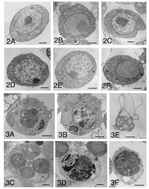
This is a small cell and most of its body contains from nucleus and cytoplasm also, around nucleus this cell has 3micrometer and normally has a big nucleus (Hirose et al,2003). Additionally, the cytoplasm in hemoblast has some organs such as mitochondria, Golgi bodies, rough endoplasmic reticulum and a large number of ribosomes and the job if these ribosomes are active biosynthesis (Hirose et al,2003). Moreover, Hemoblast has few vacuoles, whereas, some of them has small granules (Hirose et al,2003).
Phagocyte:
Characteristic of phygocytes comes by phagosomes and pseudopodia, also, the count and size of phagosomes are changeable (Hirose et al,2003). Although, phagocytes have usually other hemocytes in phagosomes, but, sometimes two or more have eaten from hemocytes by phagocytes (Hirose et al,2003). Phagosomes actually has two situations in the cell, firstly, when the size of phagosomes is small from the total of cell’s size that’s mean the total volume is small usually, has several long pseudopodia (Hirose et al,2003). On the other hand, when the phagosomes are a big size and take the most of the cell’s size that caused the cytoplasm is often thin layer around the engulfed materials and pseudopodia (Hirose et al,2003). The genus which has this phagocyte B. fuscus (Hirose et al,2003).
Granulocyte:
This type of cells has several circles or elliptical granules with a different size and different feature, also, these granules cells are different based on the species (Hirose et al,2003). For example, in the genus called B.primigenus the granulocytes in around sharp and 0.4micrometers in diameter (Hirose et al,2003). In other species such as B.scalaris and B. schlosseri nearly 0.7micrometers in diameter (Hirose et al,2003). In some granules with elliptical feature founded in B. schosseri the size is depend on the species and it is different between different species about 0.5 micrometer in diameter in B.violaceus (Hirose et al,2003). Additionally, 0.1micrometers in B. sexiens and B. fuscus and about 1.5 micro meters B. delicatus and B. simodensis, moreover, in B. lentus is round granules with 1.8 micro meters (Hirose et al,2003). In specie B. scalaris granulocytes normally have a rod haped and the granule is large and has tessellated substructure (Hirose et al,2003). The previous species have similarities substructure in large granule, tunic cells and the type of free cell distributed in the tunic (Hirose et al,2003).
Morula cell:
“Morula cells are round and contain several vacuoles that are filled with electron dense materials” (Hirose et al,2003). Even in some species like B. primigenus, the size and number of these cells are changeable (Hirose et al,2003). In the last species vacuolar insides on some morula shaped and hemocytes are much “more electron dense than usual morula cells” (Hirose et al,2003). “They are abundantly found in the mantle of upper part of the zooids. In the live specimens, they are dark purple contributing to the colony color of this species and are referred as pigmentary morula cells in this report. In B. schlosseri, each vacuole contains an electron dense sphere, and there is a clear space around the sphere. Whereas this type of morula cells is sometimes found in the other botryllid species, most of the morula cells are this type in B. schlosseri. Some morula cells are univacuolar in B. fuscus” (Hirose et al,2003).
Pigment cell:
This cell is a huge also, the circle shape in this cell without psedupoda but these cells have few more than one large vacuoles thrusting and the nuclei and the cytoplasm in the outer layer (Hirose et al,2003). In this cell the vacuoles have several pigment granules without limited number also, the granules differ in shape and size, in addition, every pigment cell has one type of the granules (Hirose et al,2003). In fact, could be some differences of the granules type which will explain, such as the ultrastructure of nephrocytes essentially not has any different from the other pigment cells (Hirose et al,2003). While the structure of granule has sometimes different from other pigment granula (Hirose et al,2003). The pigment cells are usually originate in B.sexiens, also, these cells have a large vacuole without granules, “Although they may be precursors of pigment cells, hemocytes of similar morphology are rarely found in the other species examined here. Therefore, this hemocyte type is temporally referred as large-vacuole cell” (Hirose et al,2003). In B.lentus some vacuolated hemocytes have moderately electron dense materials also, they are a similarity with morula cells in morphology side, however, these cells have a different characteristic such as the electron density in vacuolar contents are lower than morula cells moreover, the usually the vacuolar membranes obscure (Hirose et al,2003). In botryllid ascidianse especially in the colony system the young oocytes identify as a circulate with blood in the vascular system (Hirose et al,2003). According to Hirose et al (2003), the hemocytes are much smaller than oocytes. In the centre of oocytes has a circle nucleus and also a big important nucleolus (Hirose et al,2003). The oocytes are enclosed by the primary follicle cells, additionally, the follicle cells have several nucleolus and this caused to make the attached is difficult, whereas the attached loosed by this cells (Hirose et al,2003). In the next table according to Hirose et al (2003), comparison between different type of hemocytes cells and some genera in ascidiacea class.


To sum up, this subphylum Urochordate has more than one thousand specious. This subphylum follows the chordate phylum with the two another subphylum first one is cephalochordate and second is the subphylum vertebrate. The subphylum Urochordate is the biggest group in the marine invertebrate because it has more than 3000 species. In addition, the subphylum Urochordata has two orders and several suborders for each also, has a great number of families and subfamilies and that caused to contain more than 3000 species. Although has a massive number of species but it has a limit hemocytes group from 5-9 types of hemocytes cells. Moreover, the ascidians group has a network cell but is very sensitive any heavy materials such as EDTA. In addition, the network cells related directly with muscle. In fact, this subphylum is a significant group in the kingdom Animalia.
References:
Ali, H. A. J., & Tamilselvi, M. (2016). Classification of Ascidians. In Ascidians in Coastal Water (pp. 13-18). Springer International Publishing.
Hirose, E. (2009). Ascidian tunic cells: morphology and functional diversity of free cells outside the epidermis. Invertebrate Biology, 128(1), 83-96.
Hirose, E., Shirae, M., & Saito, Y. (2003). Ultrastructures and classification of circulating hemocytes in 9 botryllid ascidians (Chordata: Ascidiacea). Zoological science, 20(5), 647-656.
McHenry, M. J., & Patek, S. N. (2004). The evolution of larval morphology and swimming performance in ascidians. Evolution, 58(6), 1209-1224.
Nishino, A., & Satoh, N. (2001). The simple tail of chordates: phylogenetic significance of appendicularians. Genesis, 29(1), 36-45.
Satoh, N., Rokhsar, D., & Nishikawa, T. (2014, November). Chordate evolution and the three-phylum system. In Proc. R. Soc. B (Vol. 281, No. 1794, p. 20141729). The Royal Society.


- 24/7 Customer Support
- 100% Customer Satisfaction
- No Privacy Violation
- Quick Services
- Subject Experts