Understanding the Components and Functions of the Respiratory
Structured essay:
Describing the components of respiratory system and explaining their function in the ventilation and gas exchange: (1.2)
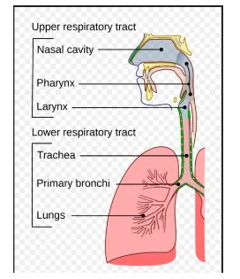
Respiratory system contains three major components:
Upper airways
Lower airways
Pleura
Upper airways:
The upper airways of a respiratory system consists of nose, nasal cavity, mouth, and pharynx. Larynx and trachea (Azimi et al. 2017).
Nasal cavity:
This is the beginning of the upper respiratory tract which opens towards the face through two nares anteriorly. Posteriorly the nasal cavity opens to the nasopharynx by two choanae (Peate, 2018).
The roof of the nasal cavity is made up of nasal and frontal bones anteriorly (Gordon and Reed, 2020).
The anterior part and nares of nasal cavity consist of sebaceous glands as well as hair follicle which prevent the entry of harmful particle into the nose.
Nose:
Nasal cavity and nose form the upper portion of the respiratory tract.
Nose contains two rounded as well as oval holes which are termed as nostrils.
Through nostril air enters into the nose then passes to the nasal cavity.
Pharynx:
It is the funnel shaped and muscular structure that is attached to the nasal cavity and paranasal sinus posteriorly and larynx anteriorly (Longmore et al. 2019).
Pharynx consists of three major parts such as nasopharynx, laryngopharynx and oropharynx
Nasopharynx:
The superior-most and the first part of pharynx which is attached the nasal cavity posteriorly (Cupello et al. 2017).

This part consists of ciliated epithelium which enables this part to receive the fresh air from nasal cavity and push the air towards larynx. While eating foods, this part remains closed thereby restricting the entry of food into respiratory tract.
Oropharynx:
It is attached posteriorly to the oral cavity
It is lined up with highly protective non-keratinizing, squamous stratified epithelium which enables this part to pass both the food from oral cavity and air from the nasal cavity towards tire destination (Longmore et al. 2019).
Laryngopharynx:
The inferior-most part of larynx which divides the respiratory and digestive tract into two separate system (Bains et al. 2020).
This part is attached to larynx anteriorly thereby supplying air towards larynx.
The hollow and complex structure that opens anteriorly to the esophagus and posteriorly to pharynx (Zaccone et al. 2018).
It is protected by the squamous epithelium and lined up with squamous stratified epithelium.
Lower respiratory tract:
Lower respiratory tract consists of trachea, bronchi, bronchioles, lungs and alveoli.
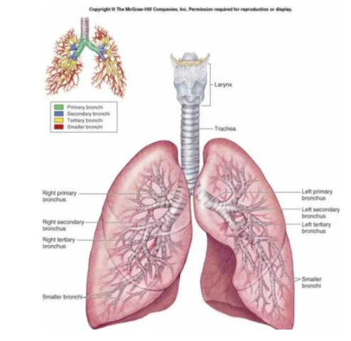
Trachea:
Pipe like flexible structure which opens anteriorly to larynx and posteriorly to bronchi.
It consists of small rings of cartilaginous tissues which support and surround this structure (Bains et al. 2020). The cartilaginous rings protect this structure from collapsing while changing the air pressure inside. This structure is specially made for letting the air to pass through the trachea and bronchioles and then goes to lungs.
Bronchi
The trachea is divided into two branches which are known as left and right bronchi. The left and right bronchi enter into the left and right lungs respectively (Gordon and Reed, 2020).
Bronchi is supported by cartilaginous rings like trachea.
Bronchioles:
After entering into lungs, each left and right bronchi is further subdivided into many breaches which are known as bronchioles ((Zaccone et al. 2018).
Lungs:
This is the primary respiratory organ in human respiratory system which plays crucial roles in gas exchange and ventilation (Depner et al. 2017).)
The paired structure that are situated into the thoracic cavity
Inside lungs there are many branches of bronchi which are subdivided into bronchioles. These bronchioles form tiny air sacs or alveoli at end.

Alveoli:
These are the tiny air sacs that are formed at the need of the bronchiole. Alveoli is surrounded by many blood vessels and capillaries (Garcia Parraga et al. 2018).
Alveoli play major roles in gas exchange and blood oxygenation.
Functions ventilation and gaseous exchange:
Pulmonary ventilation is the process of gaseous exchange in which the fresh air (oxygen) enters into lungs via different upper and; lower respiratory organs and CO2 exits from the body to the external environment (Park et al. 2020).

Inhalation:
In halation is the process of entering oxygen of fresh air from outside the body into the nose and then transported finally to the lungs (Azimi et al. 2017).
During this process, the air from outside enters into the nasal cavity through nose (Gordon and Reed, 2020). The ciliated epithelium in the nasal cavity plays crucial roles in restricting the entry of harmful articles into the nose hereby ensuring that only fresh air enters into the nasal cavity.
From the nasal cavity, the air then passes to the pharynx (Longmore et al. 2019). This tubular structure is specially made up of stratified ciliated epithelium that swipes the air from laryngopharynx to the larynx.
After entering into larynx, the air then passes towards the lower respiratory tract and enters into the trachea and then to bronchi.
From bronchi the air enters into lungs and then reached the alveoli or air sacs. Alveoli or air sacs is surrounded by network of blood capillaries (Cupello et al. 2017).
Alveoli plays crucial roles in gaseous exchange. After the fresh air (oxygen) enters into alveoli, it then passes to the blood surrounding blood vessels through diffusion. Similarly the CO2 from the deoxygenated blood of blood vessels enters into alveoli through diffusion which then exist from the body (Zaccone et al. 2018).
The oxygen that enters into the blood vessels in then transported through blood to heart and then to entire body parts.
Exhalation:
Exhalation is the process in which CO2 is breathed out of the body from lungs. From pulmonary artery of heart, deoxygenated blood is transported to the blood vessels that surrounds alveoli (Zaccone et al. 2018).
Diffusion takes place and CO2 in the blood inside the capillaries enters into alveoli.
From alveoli the CO2 is then transported through bronchi, trachea, and larynx and then finally CO2 is exhaled out from the body to the outside environment.
Explaining how respiratory system interacts with the cardiovascular system for maintaining proper body functions: (1.3)
Both the cardiovascular system and respiratory system work together to ensure that all the body cells and tissues receive enough oxygen to perform cellular activities (Cupello et al. 2017). Respiratory system plays crucial roles in inhaling sufficient oxygen from the external environment. The oxygen is then transported to the blood from alveoli through gaseous exchange and then the oxygen is transported to heart (Park et al. 2020). From heart. While oxygen enters into heart the cardiovascular system plays crucial roles in sending oxygenated blood from heart to different body cells thereby enabling to perform normal body functions. Both the cardiovascular and respiratory system work together in the following ways:
The respiratory system:
This system consists of larynx, trachea, two bronchi, bronchioles, lungs and alveoli. Respiration is the process of inhalation and exhalation of oxygen (Gordon and Reed, 2020). While air is breathed in the body it is transported through larynx, trachea, bronchi and bronchioles. Finally the air enters into lungs and then alveoli. The outer wall of alveoli is surrounded by dense capillary network which allows gaseous exchange to be taken place thorough the alveolar wall (Zaccone et al. 2018).). By diffusion the oxygen within the alveoli enters into the surrounding capillaries and mixed up with the blood inside. The blood is then termed as oxygenated blood. Then the oxygenated blood is then transported to heart for further process which is done by cardio vascular system. For those requiring assistance with their healthcare dissertation, seeking healthcare dissertation help can be valuable for understanding complex physiological processes like this one and integrating them into a comprehensive analysis.
Cardiovascular system:

Cardiovascular system consists of heart and blood vessels that play crucial roles in carrying oxygenated blood from respiratory system thereby supplying oxygenated blood to different body parts (Bains et al. 2020). Heart is located at junction of the circulatory and respiratory system thereby maintaining connection between these two systems. The heart consist of two atria and two ventricles. During circulation, the deoxygenated blood enters into the right atrium from veins (superior and inferior vena cava) (Gordon and Reed, 2020). Then the relaxation of the cardiac muscles occurs which pushes the blood to enters into the right ventricle. Then from the right ventricles the deoxygenated blood is pushed into the pulmonary artery. Pulmonary arteries carry the deoxygenated blood into the blood vessels that surround alveoli (Zaccone et al. 2018). At this moment diffusion takes place which enables the CO2 from the deoxygenated blood inside blood vessels surrounding the alveoli enters into alveoli and then CO2 passes through lower and upper respiratory tract to outside the body.

After diffusion this oxygen enters into the blood vessels that surrounds alveoli. This process is termed as blood oxygenation (Longmore et al. 2019). In this process the oxygenated blood is then transported from the blood vessels the surrounds the alveoli to the pulmonary vein. Pulmonary vein is the only vein that carries the oxygenated blood. From pulmonary vein the oxygenated blood is then enters into the left atrium. Then from the left atrium the oxygenated blood then enters into the left ventricle through aortic valve due to relaxation of cardiac muscle (Cupello et al. 2017). Finally the oxygenated blood is pushed into the aorta which carry blood to different body parts. In this way the oxygenated blood is transported to all the body parts thereby enabling the cell to perform the cellular respiration.
Reference list:
Azimi, H., Gilakjani, S.S., Bouchard, M., Bennett, S., Goubran, R.A. and Knoefel, F., 2017, May. Breathing signal combining for respiration rate estimation in smart beds. In 2017 IEEE International Symposium on Medical Measurements and Applications (MeMeA) (pp. 303-307). IEEE.
Bains, K.N.S., Kashyap, S. and Lappin, S.L., 2020. Anatomy, Thorax, Diaphragm. StatPearls [Internet].
Cupello, C., Meunier, F.J., Herbin, M., Clément, G. and Brito, P.M., 2017. Lung anatomy and histology of the extant coelacanth shed light on the loss of air-breathing during deep-water adaptation in actinistians. Royal Society open science, 4(3), p.161030.
Depner, M., Ege, M.J., Cox, M.J., Dwyer, S., Walker, A.W., Birzele, L.T., Genuneit, J., Horak, E., Braun-Fahrländer, C., Danielewicz, H. and Maier, R.M., 2017. Bacterial microbiota of the upper respiratory tract and childhood asthma. Journal of Allergy and Clinical Immunology, 139(3), pp.826-834.
Garcia Parraga, D., Moore, M. and Fahlman, A., 2018. Pulmonary ventilation–perfusion mismatch: a novel hypothesis for how diving vertebrates may avoid the bends. Proceedings of the Royal Society B: Biological Sciences, 285(1877), p.20180482.
Gordon, K.E. and Reed, O., 2020. The role of the pelvic floor in respiration: a multidisciplinary literature review. Journal of Voice, 34(2), pp.243-249.
Katzenberg, M.A. and Grauer, A.L. eds., 2018. Biological anthropology of the human skeleton. John Wiley & Sons.
Longmore, S.K., Lui, G.Y., Naik, G., Breen, P.P., Jalaludin, B. and Gargiulo, G.D., 2019. A comparison of reflective photoplethysmography for detection of heart rate, blood oxygen saturation, and respiration rate at various anatomical locations. Sensors, 19(8), p.1874.
Park, A., Song, Y., Yi, E., Duy Nguyen, B.T., Han, D., Sohn, E., Park, Y., Jung, J., Lee, Y.M., Cho, Y.H. and Kim, J.F., 2020. Blood Oxygenation Using Fluoropolymer-Based Artificial Lung Membranes. ACS Biomaterials Science & Engineering.
Peate, I., 2018. Anatomy and physiology, 10. The respiratory system. British Journal of Healthcare Assistants, 12(4), pp.178-181.
Wang, L., Zhang, X., He, Y., Wang, Y., Zhong, W., Mequanint, K., Qiu, X. and Xing, M., 2019. Ultralight conductive and elastic aerogel for skeletal muscle atrophy regeneration. Advanced Functional Materials, 29(1), p.1806200.
Zaccone, G., Lauriano, E.R., Capillo, G. and Kuciel, M., 2018. Air-breathing in fish: Air-breathing organs and control of respiration: Nerves and neurotransmitters in the air-breathing organs and the skin. Acta histochemica, 120(7), pp.630-641.
Continue your exploration of Understanding Play Therapy as an Intervention for Autistic Children with our related content.


- 24/7 Customer Support
- 100% Customer Satisfaction
- No Privacy Violation
- Quick Services
- Subject Experts